Laser Gum Surgery in Sacramento, CA Tooth & Bone Loss
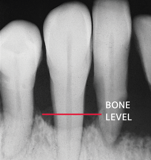
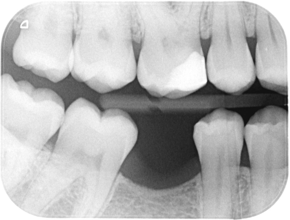
The range of normal bone levels are when the crest of the alveolar ridge is 0.5 to 2.0 mm apical to the cemento-enamel junction. In the anterior region, the crest of the alveolar ridge may have a pointed appearance due to smaller interproximal spacing between the teeth. In the posterior region, the crest of the alveolar ridge will be flatter.
PREVENTING PERIODONTAL DISEASE Battlefield Modern Dentistry
Low bone mineral density and osteoporosis. Osteoporosis is a disease characterised by low bone mass and microarchitectural deterioration of bone tissue leading to enhanced bone fragility and a consequent increase of fracture risk ().The outer, cortical, layers of the bone become thinner while the trabecular structure becomes more open ().There is a progressive reduction in bone mineral density.
Bone Loss Just Wright Dental

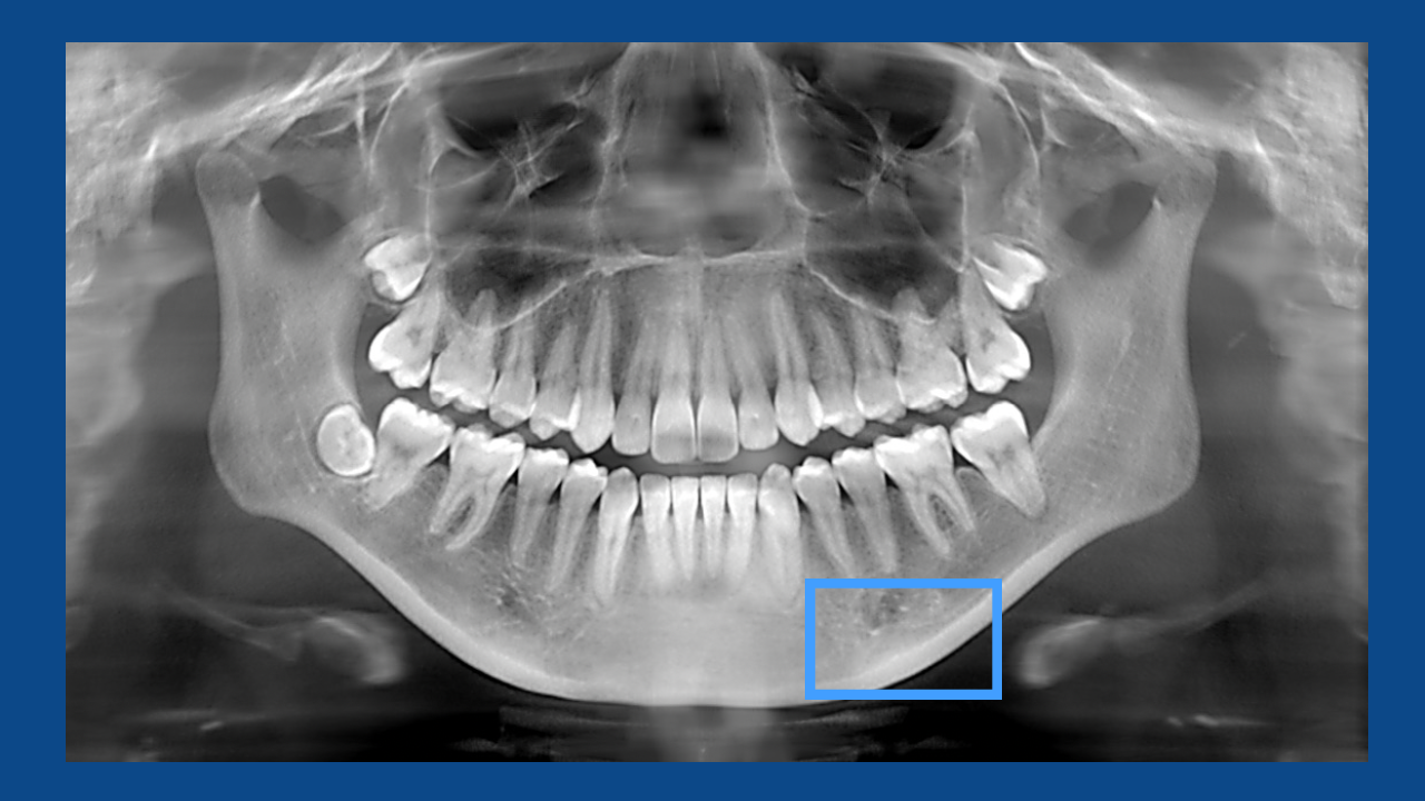
Background: Intraoral radiographs can aid in formulating a more accurate diagnosis of periodontal disease. However, it must be considered whether a comparable amount of information can be obtained with modern panoramic radiographs. The aim of this study was to determine to what degree the diagnosable amount of bone loss in patients with aggressive periodontitis or severe chronic periodontitis.
Can you Still Have Dental Implants if You Have Severe Bone Loss?

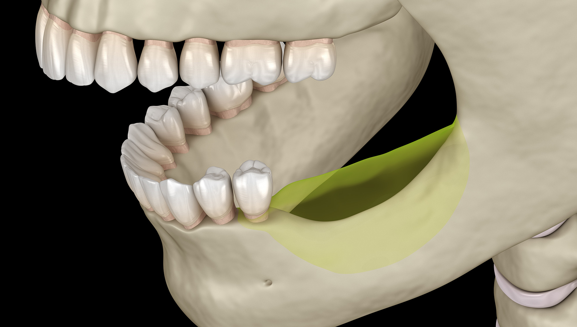
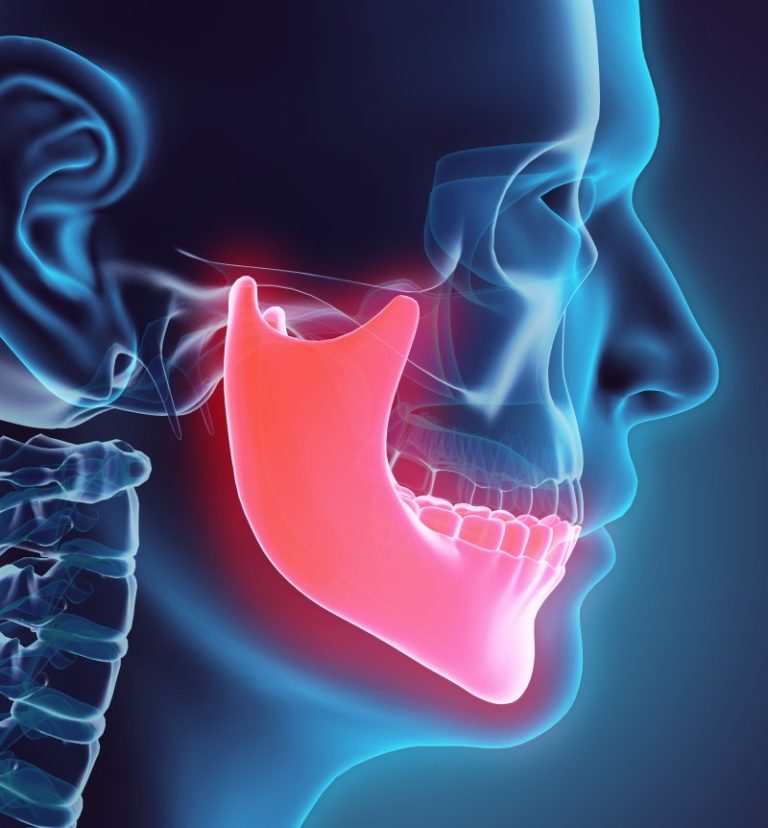
When you think of oral health issues, cavities and bad breath are usually top of mind. But there's another common (and quite serious) condition that can affect anybody, even those with perfect pearly whites: bone loss. Technically speaking, teeth aren't bones.When you hear about "bone loss in teeth," this is actually referring to the alveolar bone that surrounds and supports your teeth.
View Image

Abstract. Osteoporosis is a severe skeletal disease that leads to bone fractures, even disability, if it remains undetected. However, osteoporosis remains frequently unnoticed until a fracture occurs. It is possible for dental practitioners to screen patients at risk of osteoporosis and refer them for an osteoporosis evaluation.
4 loose teeth and tenderness on upper jaw after orthognathic surgery. Xrays show black spots on

The gum tissue can appear at a healthy level even with a significant amount of bone loss. Therefore, dental radiographs are needed to assess what the bone height is and whether there is any loss of the bone support for the teeth.
Replacement Teeth With Advanced Periodontal Disease PPIC
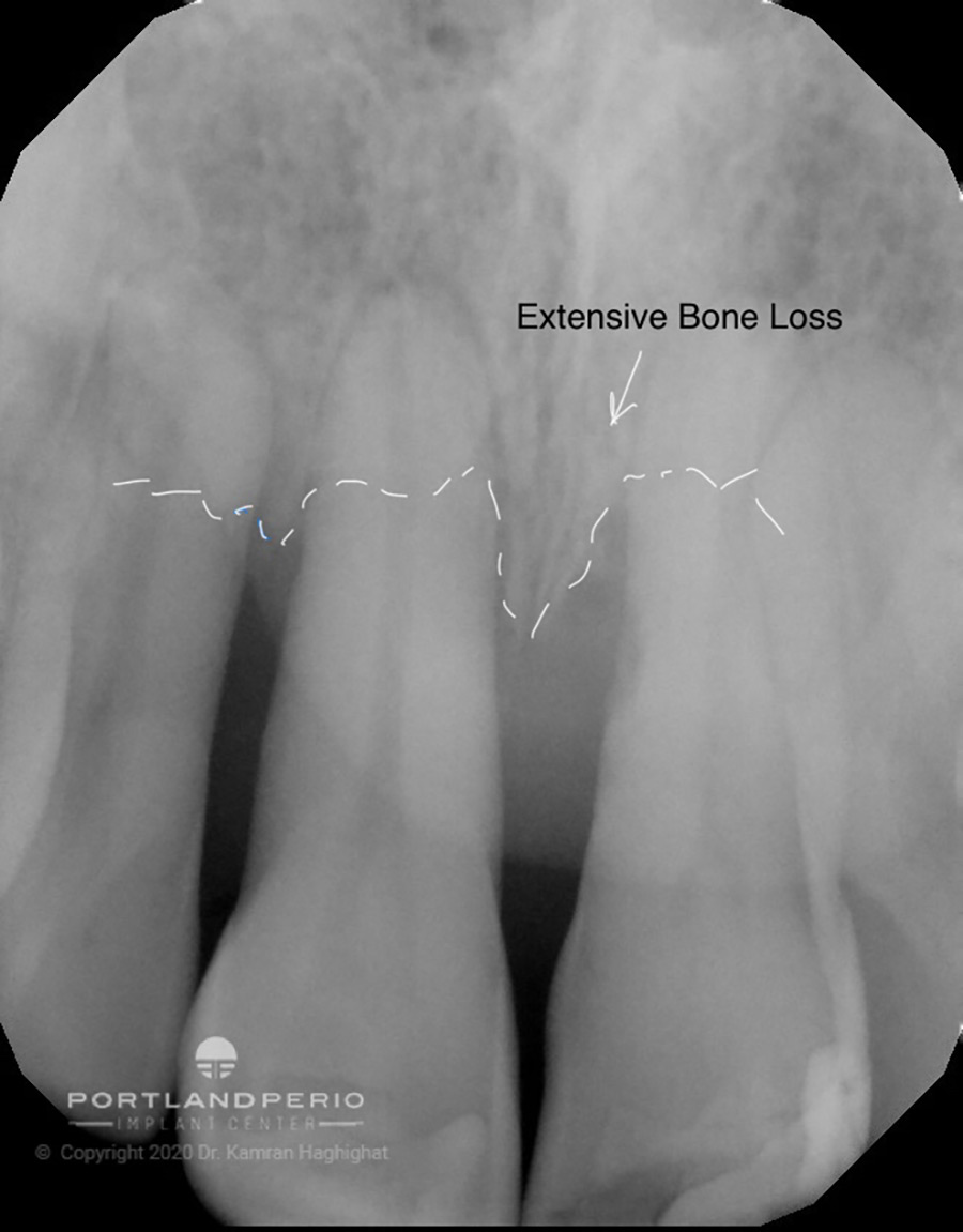
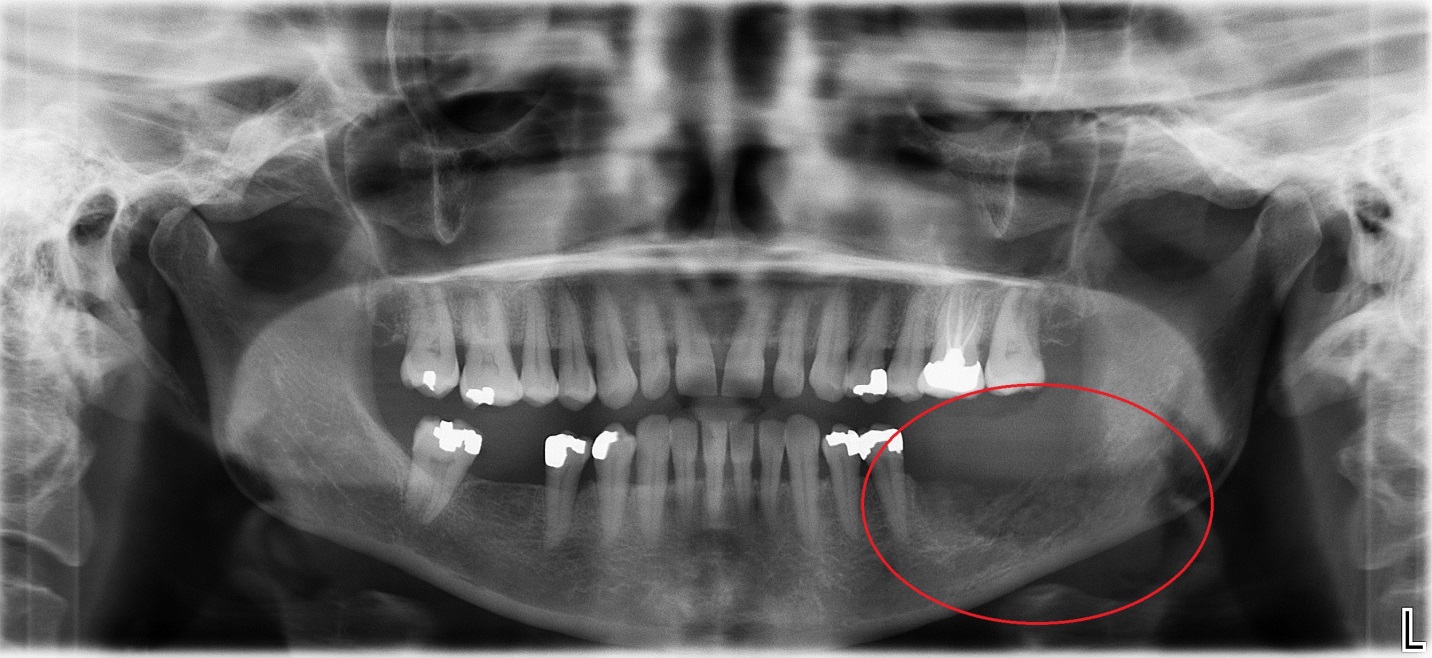
Yes, x-rays can help show severe bone loss in teeth, which is a sign of periodontal disease. When caught early, this serious form of gum disease can be treated and managed. That's why Reveal Diagnostics offers convenient imaging centers across the Bay Area that can take the necessary x-rays to help diagnose periodontal disease.
AI Detects Osteoporosis With High Accuracy By Analyzing Dental XRays
Injury and irritation of the facial bones can negatively affect bone cell activity and reduce bone regrowth. For example, a bad fall, car accident, or sporting injury could damage the bones in the face, leading to bone loss. 4. Continually grinding your teeth ( bruxism) can also stress the jaw and traumatize the teeth, resulting in bone resorption.
Bone Loss in Wakefield Missing Teeth Schumacher Dental
Radiographic Changes seen in Periodontal Disease. Periodontal disease causes inflammatory lesions in the marginal bone Both osteoblastic and osteoclastic activity is seen Osteoclastic activity will cause changes in the morphology of the crestal bone Initial response is destruction of bone. Chronic lesions will show some osteosclerosis.
Why Do I Need Xrays? Your Prescott Dentist Hicks Dental Group

In contrast to two-dimensional planar images, a measuring point is hardly repeatedly determined in a CBCT image when alveolar bone loss is assessed. Thus, the aim of the present study was to propose a six-site measuring method, which is closely related to anatomical structure, for the evaluation of alveolar bone loss in CBCT images.
Pin on Dental Implants

1. Introduction. Periodontitis is a prevalent dental health problem and can be classified as a major global challenge that affects developed and developing countries [1,2,3].Triggered by bacterial colonization of the root surface, the host's immune system reacts with inflammatory processes to the microbial transition from a symbiotic bacterial environment to that of dysbiotic pathogens.
Why your Dentist takes Xrays... Brucegate Dental Practice
The dental x-ray radiograph shows the extent of the tooth damage. The #9 tooth root was then extracted from under the tooth crown to allow the gum and bone to heal eight months before the completion of her dental braces. 3) & 4) This rootless tooth crown #9 is being held in place only by the orthodontic bracket and wire.
XRay of Dark Resorption in Jaw Bone
The recent change in classification of periodontal and peri-implant diseases includes objective evaluation of intra-oral radiographs and quantification of bone loss for disease staging and grading. Assessment of the progression of periodontal disease requires deduction of bone loss longitudinally, and its interpretation as (1) a percentage in relation to tooth root and (2) as a function of the.
Fall Creek Dental Blog The Deal with Dental XRays

Application error: a client-side exception has occurred (see the browser console for more information). Coming in September: Interpreting endodontic X-rays will be the subject of the next article in Dr. Bellows' radiography series.
What You Should Know About Dental XRays Nicole Mermet, DMD Dentist

Radiographs are an integral component of a periodontal assessment for those with clinical evidence of periodontal destruction. A close consideration of the current approach to periodontal diagnosis compatible with the current classification of periodontal diseases reveals that radiographs only inform with respect to diagnosis for a small proportion of conditions.
Bone Loss in Teeth Causes, Prevention & Treatment 1311 Jackson Ave Dental Dentist in Long

This week is all about the basics of horizontal bone loss and vertical bone defects on radiographs. Horizontal Bone Loss. When identifying horizontal bone loss you must first go through the same steps of evaluating normal bone appearances (last weeks post).. The two lines (adjacent cemento-enamel junctions and crest of the alveolar ridge) will still be parallel.